function
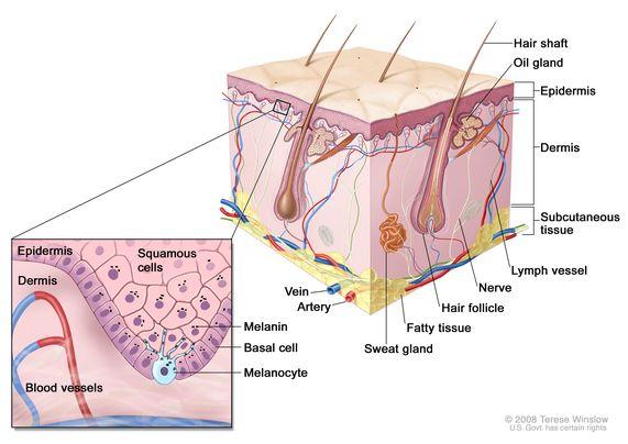
melanocytes produced melanin sun radiation prevents skin damage caused by cell chromosome. Melanoma cells can synthesize and secrete melanin, thus a gland cells. However, melanin biosynthesis very complicated by the color bodies (immature melanin) tyrosine - tyrosinase formed by the reaction.
in normal human epidermis, a melanocyte about 40 can take into account the keratinocytes, the epidermis forming units called melanocytes. Color of the skin from the keratinocytes storing melanin in the cells. In general, more people are storing melanin skin color deeper, more protected, away from sun radiation.
identified melanocytes
Identification of melanocytes (melanocyte, MC) cultured in vitro, is substantially the following.
Masson-Fontana silver by ammonia
melanin granules and is mainly used to identify argyrophilic grain. Melanin is a non-fat-soluble, non-hemoglobin of pigment particles, dark brown, are present in certain parts of the skin, hair, retina, iris, and central nervous system. Argyrophilic grain distribution carcinoid. After staining and Argyrophilic grain melanin showed black, pink nuclei, cytoplasm pale red.

Dopa staining
is the synthesis of melanin tyrosine as substrate, under the action of tyrosinase, into dopa after hydroxylated, further oxidized to multiple Pakistan quinone, further become melanin. Addition of exogenous levodopa tyrosinase-specific, cell culture in the presence of MC, will be under the action of tyrosinase more synthetic pigment particles under the microscope the cells appear black.
SEM direct observation of cell structure wherein melanosomes
no tension within the filament and the electron microscope observation desmosomes MC, visibility characteristic of melanosomes (melano- some), tyrosinase-containing organelle. In the next cell nucleus I, II stage of bodies melanin is more common in the surrounding cytoplasm of mature III, IV stage of bodies melanin common, visible bodies mature stage IV melanosomes in the dendrites projecting's are arranged on both sides of dendrite, the apical dendrite delivered to. In addition to the melanosome structural MC visible cytoplasm rich in mitochondria and abundant endoplasmic reticulum and ribosomes.
expression in cells was detected by immunohistochemistry of various proteins
Currently, there are several commonly used marker:
①S100 Protein: S100 protein is a calcium binding protein of low molecular weight group, a relative molecular weight of 10x10 3 ~ 12x10 3 , which amino acid sequence is highly conserved in vertebrates, and calmodulin having high homology . At present, were found to have 20 kinds of structure and function similar to exist in different parts can be adjusted intracellular and extracellular Ca 2 + S100 of proteins, including S100A1 ~ S100A13, S100B, S100P, calgranulin C, calcium binding protein 3 and the like. Wherein, S100B in glial cells mainly distributed in the central nervous system and peripheral nervous system, and Schwann cells, and some neuronal cells, melanocytes, chondrocytes, and adipocytes, while S100A4 mainly distributed in fibroblasts, mainly S100A7 distributed in the epithelial cells.
② ganglioside GD3: gangliosides (gangliosides, GLS) are a class of glycosphingolipids containing sialic acid, because it is found in the first named ganglion cells. Depending on the purpose of its sialic acid residues contained, it can be divided into GM, GDGT GQ, and the like. Each ganglioside also based on the size of the mobility of the thin layer chromatography Rf value subdivided several subclasses, such as GD1, GD2, GD3 and the like. Cross-GM3 antigens is called melanoma, but GD3 than GM3 more specific, both in the normal ratio of GM3 MC: GD3 to 18: 1, in melanoma GM3: GD3 1:15.
③ melanosomes specific antigen melanosomes 1 and -5 specific antigen: Studies show that melanosomes specific antigen (humanmelanosomespecificantigens, HMSA) -1 HMSA-5 and melanin tumor staining positive rate was 60% and 69%, respectively. HMSA-1 is mainly expressed in the non-pigmented tumors and salivary gland tumors. Epithelium, which is mainly used marker of malignancy MC, MC normal skin cultures were negative. HMSA-5 recognizes a glycoprotein of 69 ~ 73kDa, appeared in all forms of human MC, may be lost in the future of the maturation process. Thus, generally with HMSA-5 labeled normal MC, MC staining in vitro. DHMB45 (anti Gp100): Gp100 is a glycoprotein of 100kDa, NKI / beteb, HMB-45, HMB-50 and ME20 this antigen are identified. Wherein, HMB-45 mAb identify immature melanosomes, MC expressed in neural crest tumors and tumor, normal breast epithelium, and some breast, sweat glands, and - a number of sweat glands and other tumors also expressed than in the diagnosis of malignant melanoma aspect S100 or NKI / C3 more specific. Thus, HMB-45 labeled with malignant MC, MC normal culture negative staining.
⑤ tyrosinase (of TYR) and tyrosinase-related protein 1,2 (TRP1 and TRP2): These are the enzymes of melanogenesis MC, MC are also used to identify.
⑥Melan-A (- MC differentiation marker species): Melan-A belongs to the expression product of the gene, called MART-1 (T cell recognition of melanoma antigens), from melanoma cell lines cloned from. It subcellular site is uncertain, but is considered and melanosomes and associated endoplasmic reticulum, aberrant expression of this gene in the MC skin, retina, and malignant melanoma, in addition to MC, the expression of the gene can be vascular muscle cells lipoma. Two monoclonal antibodies are bred A103 and M2-7C10. Both antibodies suitable for use in formaldehyde, paraffin-embedded tissue Chou, A103 the most widely used material in the towel applied research organization, M2-7C10 mainly for research cytology specimens.
⑦CK16: Some scholars have studied the expression of cytokeratin in the case of the MC, the MC were found in all of CK16 expression, but not expression CK1, CK5, CK8, CK10, CK14, and CK16 and often played. co-expressed on keratinocytes CK6 also no expression.
⑧ Other: MC many specific antigen receptors on the surface or constantly found, as Sox10. Sox10 is a neural tube (crest) transcription factor, Schwann cells and the maintenance of the MC differentiation, maturation and function, play an important role. Sox10 antibodies can be applied to a variety of neural crest-derived tumors, mesenchymal and epithelial tumors and normal tissues. 97% and 49% of melanoma malignant peripheral nerve sheath tumors expression of Sox 10, and located in the nucleus, therefore, considered to be a Schwann cell MC and a broad spectrum marker. In addition, CD40 and Toll-like receptors 2,4,7,9 like.
Scientific Applications
Research melanoma cells improves the understanding and mastering melanin production mechanism and its regulation in the beauty and cosmetics sector can create new whitening products, may be in the medical field treatment of albinism, vitiligo, black poisonous swollen disease and so on and so on.
related diseases
medical study found that melanoma cell damage, can lead to lack of melanin, causing vitiligo main principle is: melanoma cell injury, dropped out after being damaged melanin cells can then release the antigen stimulates the production of anti-melanoma cell antibodies more, so that more melanin cells are destroyed, resulting in a vicious cycle, resulting in a large number of melanocytes inactivation, thereby forming a skin surface shapes, size is not a spot, which is vitiligo.
high content of food
formedmelanin formation tyrosine dopa under the action of tyrosinase, and thus oxidation of melanin. The tyrosinase present in our usual foods, to enhance its activity by binding to copper, zinc, iron and other trace elements. Copper, zinc, iron rich foods: such as organ meats, meat, eggs, food black (black rice, black beans, black fungus, black sesame, etc.), seaweed and mushrooms ......
